Managing Fuchs Dystrophy: From Diagnosis To Treatment And Beyond
Fuchs endothelial corneal dystrophy, commonly known as Fuchs dystrophy, is a progressive eye condition that affects the cornea, which is the clear, dome-shaped outer surface of the eye. This disorder primarily affects the endothelial cells, which are responsible for maintaining the cornea's clarity by regulating the amount of fluid within the cornea.
In Fuchs dystrophy, the endothelial cells gradually deteriorate over time, leading to an accumulation of excess fluid within the cornea. This results in corneal swelling, known as corneal edema, and causes the cornea to become thickened and cloudy. As the condition progresses, individuals may experience symptoms such as blurred or distorted vision, glare, sensitivity to light, and discomfort or pain in the eye.
Fuchs dystrophy typically affects both eyes, although one eye may be more severely affected than the other. The condition tends to progress slowly, with symptoms often becoming noticeable in middle age or later. In advanced cases, Fuchs dystrophy can significantly impair vision and may require surgical intervention, such as corneal transplantation, to restore vision and alleviate symptoms.
The exact cause of Fuchs dystrophy is not fully understood, but it is believed to have a genetic component, with certain genetic mutations predisposing individuals to the condition. Other factors, such as age, gender (women are more commonly affected), and environmental factors, may also play a role in the development and progression of the disease.
Management of Fuchs dystrophy typically involves monitoring the progression of the condition and addressing symptoms as they arise. In the early stages, treatment may include medications or eye drops to reduce corneal swelling and alleviate symptoms. In advanced cases where vision is significantly affected, surgical options such as endothelial keratoplasty (e.g., Descemet's stripping endothelial keratoplasty or Descemet's membrane endothelial keratoplasty) or penetrating keratoplasty (corneal transplantation) may be necessary to replace the damaged endothelial cells and restore vision.
Managing
Managing Fuchs dystrophy involves a combination of lifestyle adjustments, medical interventions, and, in some cases, surgical procedures. Here are some strategies individuals with Fuchs dystrophy can employ to best manage the condition:
Regular Monitoring
Regular eye examinations with an ophthalmologist are essential for monitoring the progression of Fuchs dystrophy and assessing changes in vision and corneal health over time.
Medications and Eye Drops
In the early stages of Fuchs dystrophy, medications such as hypertonic saline ( concentrated 5% saline drops) eye drops or ointments may be prescribed to help reduce corneal swelling and alleviate symptoms such as discomfort or blurred vision.

Protective Eyewear
Protecting the eyes from irritants and excessive exposure to UV radiation can help minimise discomfort and reduce the risk of complications in individuals with Fuchs dystrophy. Wearing sunglasses outdoors and using protective eyewear in dusty or windy environments is recommended.
Maintaining Eye Health
Practising good eye hygiene, such as gently cleansing the eyelids and lashes and avoiding eye rubbing, can help prevent irritation and reduce the risk of complications in individuals with Fuchs dystrophy.
Healthy Lifestyle Choices
Adopting a healthy lifestyle that includes a balanced diet, regular exercise, adequate hydration, and sufficient sleep can support overall eye health and may help slow the progression of Fuchs dystrophy.
Surgical Interventions
In advanced cases of Fuchs dystrophy where vision is significantly affected, surgical options such as endothelial keratoplasty (e.g., Descemet's stripping endothelial keratoplasty or Descemet's membrane endothelial keratoplasty) or penetrating keratoplasty (corneal transplantation) may be necessary to replace the damaged endothelial cells and restore vision.
Educating Yourself
Learning about Fuchs dystrophy, including its causes, symptoms, and treatment options, can empower individuals to make informed decisions about their eye care and advocate for their needs with healthcare providers.
By incorporating these strategies into their daily routine and working closely with their healthcare team, individuals with Fuchs dystrophy can effectively manage the condition and maintain their vision and overall eye health.

Diagnosis
Diagnosing Fuchs dystrophy typically involves a comprehensive eye examination and specialised tests to assess the health and function of the cornea. Here are the steps involved in diagnosing Fuchs dystrophy:
Patient History: The ophthalmologist will begin by taking a detailed medical history, including any symptoms the patient may be experiencing, family history of eye conditions, and past eye surgeries or treatments.
Visual Acuity Test: A visual acuity test is performed to evaluate the clarity and sharpness of the patient's vision. This test involves reading letters or symbols on an eye chart from a specific distance.
Slit Lamp Examination: The ophthalmologist will use a slit lamp, a specialised microscope with a bright light, to examine the front structures of the eye, including the cornea. This allows for detailed visualisation of any abnormalities or changes in the corneal tissue.
Corneal Thickness Measurement: A test called pachymetry may be performed to measure the thickness of the cornea. In Fuchs dystrophy, corneal thickening due to fluid accumulation is a characteristic feature.
Endothelial Cell Density Measurement: Specular microscopy or endothelial cell counting may be used to assess the density and health of the endothelial cells, which are affected in Fuchs dystrophy. This test provides information about the number and morphology of endothelial cells per unit area of the cornea.
Guttata Assessment: The presence of guttata, which are small excrescences or bumps on the inner surface of the cornea,the presence of corneal guttata, could indicate Fuchs dystrophy. The ophthalmologist will examine the cornea for the presence of guttata during the slit lamp examination.

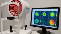
Corneal Topography: Corneal topography may be performed to map the curvature and shape of the cornea. This can help identify irregularities in corneal shape that may be indicative of Fuchs dystrophy.
Endothelial Function Tests: Specialised tests such as specular microscopy or endothelial cell function tests may be used to assess the functional capacity of the endothelial cells and their ability to maintain corneal clarity and hydration.
Genetic Testing (Optional): In some cases, genetic testing may be performed to identify specific genetic mutations associated with Fuchs dystrophy, especially in individuals with a family history of the condition.
By performing these tests and examinations, ophthalmologists can accurately diagnose Fuchs dystrophy and develop an appropriate treatment plan tailored to the individual needs of the patient.
Treatment
At My-iClinic, the treatment options offered for Fuchs dystrophy may include:
Medications
In the early stages of Fuchs dystrophy, medications such as hypertonic saline eye drops or ointments may be prescribed to help reduce corneal swelling and alleviate symptoms such as discomfort or blurred vision.
Endothelial Keratoplasty
Endothelial keratoplasty, such as Descemet's stripping endothelial keratoplasty (DSEK) or Descemet's membrane endothelial keratoplasty (DMEK), is a surgical procedure that involves replacing the damaged endothelial layer of the cornea with healthy donor tissue. This procedure aims to restore corneal clarity and improve vision in individuals with advanced Fuchs dystrophy.
Penetrating Keratoplasty (Corneal Transplant)
In cases where endothelial keratoplasty is not feasible or unsuccessful, penetrating keratoplasty may be considered. This procedure involves replacing the entire thickness of the cornea with donor tissue and is typically reserved for more severe cases of Fuchs dystrophy.
Combined Procedures
In some cases, a combination of surgical procedures may be performed to address multiple aspects of Fuchs dystrophy and optimise visual outcomes. For example, cataract surgery may be combined with endothelial keratoplasty in individuals with both Fuchs dystrophy and cataracts.
Ongoing Monitoring and Management
Following treatment, ongoing monitoring and management of Fuchs dystrophy are essential to ensure optimal outcomes and detect any potential complications or recurrence of symptoms. This may involve regular follow-up appointments with the ophthalmologist and adherence to post-operative care instructions.

These treatment options may vary depending on the severity of the condition, the patient's overall health and medical history, and their individual preferences and goals. A comprehensive evaluation and consultation with an experienced ophthalmologist in a private eye clinic can help determine the most appropriate treatment approach for each individual with Fuchs dystrophy.
Beyond
Fuchs endothelial corneal dystrophy, a progressive condition affecting the cornea, poses significant challenges to patients and clinicians alike. However, the landscape of Fuchs dystrophy management is poised for transformation with the advent of new technologies, and other groundbreaking advancements.
Diagnostics
One notable area of progress lies in the realm of diagnostics. Advanced imaging modalities, such as anterior segment optical coherence tomography (AS-OCT) and corneal topography, offer detailed insights into corneal morphology and endothelial cell function. These non-invasive techniques enable clinicians to assess disease severity, monitor progression, and tailor treatment strategies with unprecedented precision.
AI-driven algorithms
Moreover, AI-driven algorithms hold promise in enhancing diagnostic accuracy and prognostic capabilities. Machine learning algorithms trained on large datasets of corneal images can assist clinicians in early detection of Fuchs dystrophy, predicting disease progression, and optimising treatment outcomes. By harnessing the power of AI, clinicians can leverage predictive analytics to personalise patient care and intervene preemptively to mitigate disease progression.

Therapeutics
In the realm of therapeutics, novel treatment modalities are on the horizon. Emerging therapies targeting the underlying pathophysiology of Fuchs dystrophy, such as corneal endothelial cell regeneration and modulation of cellular senescence, offer potential avenues for disease modification and tissue regeneration. Additionally, advancements in tissue engineering and regenerative medicine hold promise for developing bioengineered corneal substitutes and cell-based therapies for restoring corneal clarity and function.
Innovations
Furthermore, surgical innovations are revolutionizing the landscape of corneal transplantation. Minimally invasive techniques, such as Descemet's membrane endothelial keratoplasty (DMEK), offer improved visual outcomes, faster recovery times, and reduced risk of complications compared to traditional penetrating keratoplasty. These advancements are poised to redefine the standard of care for individuals with Fuchs dystrophy, ushering in an era of precision medicine and personalised treatment approaches.
While these advancements herald a promising future for Fuchs dystrophy management, it is imperative to underscore the importance of rigorous clinical validation, long-term outcomes assessment, and equitable access to innovative therapies. Collaborative efforts between clinicians, researchers, industry stakeholders, and regulatory bodies are essential to translate scientific discoveries into tangible clinical benefits and improve the quality of life for individuals affected by Fuchs dystrophy.
In conclusion, the future of Fuchs dystrophy is characterised by a convergence of technological innovation, scientific discovery, and interdisciplinary collaboration. With continued investment in research, development, and clinical translation, we stand poised to revolutionise the paradigm of Fuchs dystrophy management and usher in an era of precision, personalised medicine for individuals worldwide.
Experience The Future Of Fuchs Dystrophy Management With My-iClinic
At My-iClinic, we are committed to providing cutting-edge, personalised solutions for individuals affected by Fuchs endothelial corneal dystrophy. Our team of expert ophthalmologists harnesses the latest advancements in diagnostic technologies, therapeutic modalities, and surgical techniques to deliver tailored treatment approaches that prioritise patient well-being and vision preservation.
With a focus on precision medicine and individualised care, My-iClinic offers comprehensive diagnostic evaluations, including advanced imaging modalities and the latest technology, to accurately assess disease severity, monitor progression, and optimise treatment strategies. Our multidisciplinary approach integrates the latest therapeutic innovations, such as corneal endothelial cell regeneration therapies and minimally invasive surgical techniques, to provide patients with the most advanced and effective treatment options available.
Don't let Fuchs dystrophy compromise your vision and quality of life. Take control of your eye health today with My-iClinic. Contact us to schedule your consultation and embark on a journey towards clearer vision and enhanced quality of life. Your vision is our priority, and we are here to guide you every step of the way.
Find out more by Speaking to our team